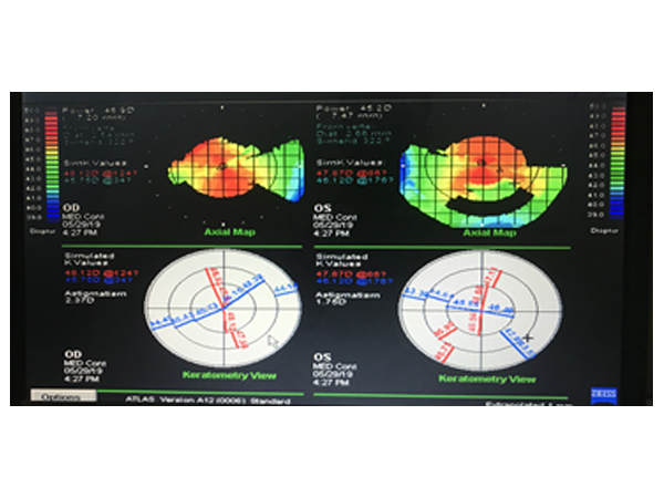
Corneal Topography
Corneal Topography is a non-invasive medical imaging technique for mapping the surface curvature of the cornea, the outer structure of the eye. Since the cornea is normally responsible for some 70% of the eye's refractive power, its topography is of critical importance in determining the quality of vision and corneal health.
Corneal topography is most commonly used for the following purposes
Refractive surgery
Keratoconus
Post surgery astigmatism
Surgical planning in cases with astigmatism
Effect of corneal and ocular surface disorders on tear film
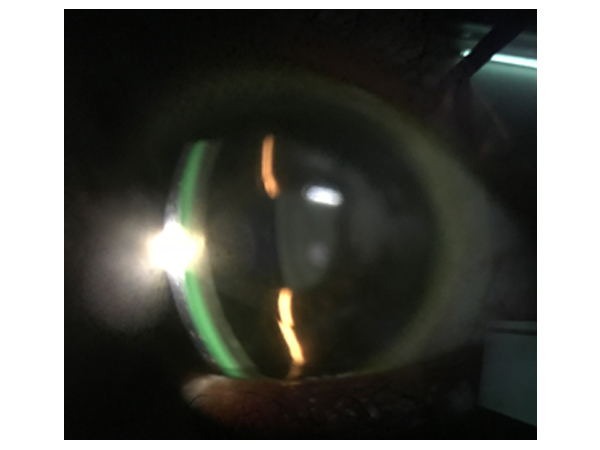
Contact lens fitting
incision placement and intrastromal ring placement in keratoconus
Pachymetry
Corneal pachymetry is the process of measuring the thickness of the cornea. A pachymeter is a device used to measure the thickness of the eye’s cornea. It is used to perform corneal pachymetry prior to refractive surgery, forscreening of Keratoconus, LRI surgery and is useful in screening for patients suspected of developing glaucoma among other uses.


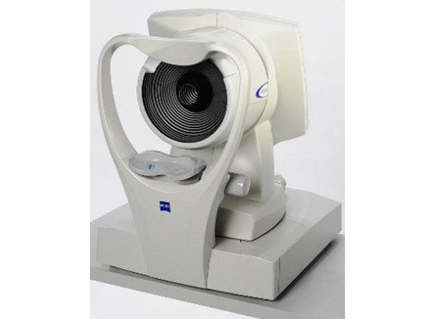
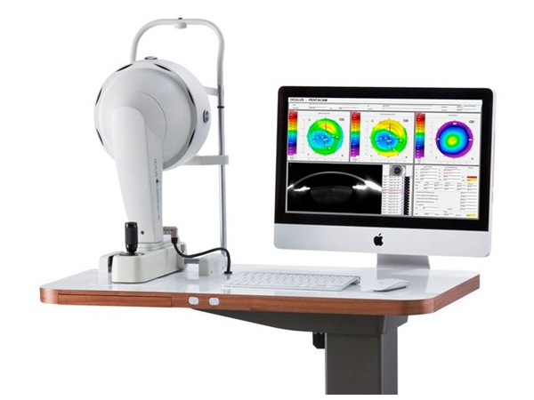
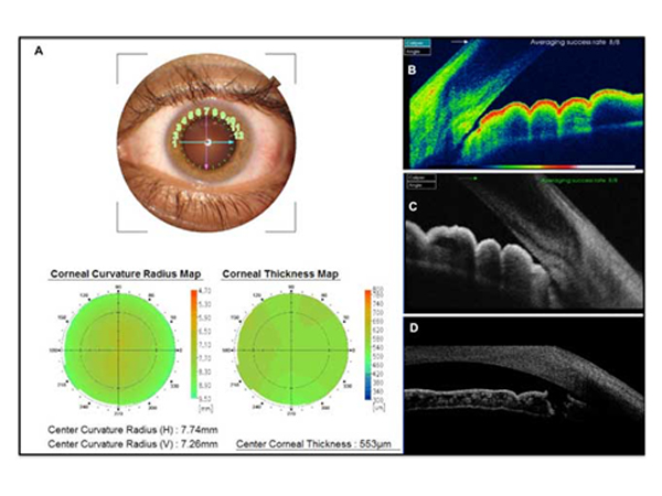
Pentacam
The Pentacam images the anterior segment of the eye by a rotating Scheimpflug camera measurement. This rotating process supplies pictures in three dimensions. The center of the cornea is measured very precisely because of this rotational imaging process. The measurement process lasts less than two seconds, and minute eye movements are captured and corrected simultaneously. By measuring 25,000 true elevation points, precise representation, repeatability and analysis are guaranteed.
The Pentacam provides 3-dimensional chamber analysis of the complete anterior segment of the eye, corneal topography of both the front and the back of the cornea, thickness measurements across the entire diameter of cornea. It also can provide cataract analysis and clearly detect corneal disorders such as Keratoconus, where laser vision correction is sometimes not appropriate.
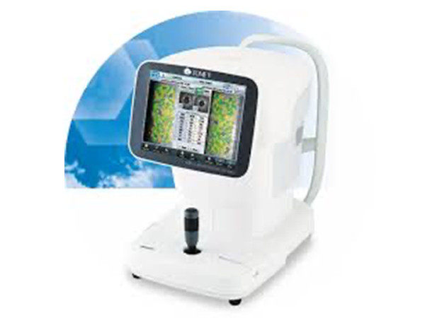
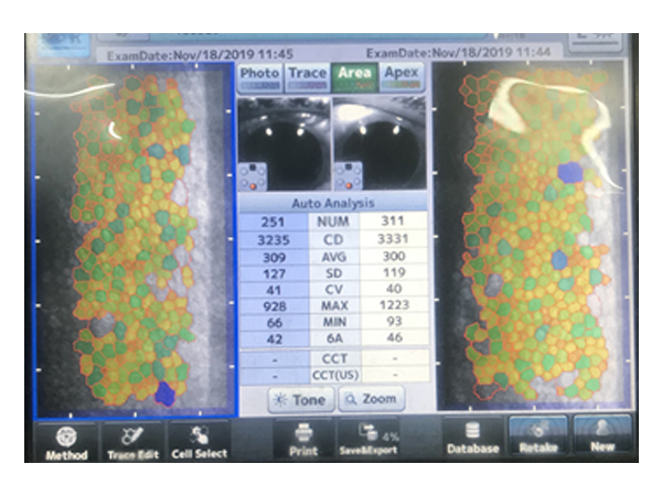
Specular Microscopy
Specular microscopy is a noninvasive photographic technique that allows you to visualize and analyze the corneal endothelium. The instrument projects light onto the cornea and captures the image that is reflected from the optical interface between the corneal endothelium and the aqueous humor.
It also measures corneal thickness
OCT for Cornea & Anterior segment
It gives corneal thickness i.e. Pachymetry
Pachymetry is important in the diagnosis of Keratoconus, Endothelial function, Descemet membrane detachment and IOP standardization
It gives assessment of angle structure
It assists in Contact Lens fitting
Staining of ocular surface
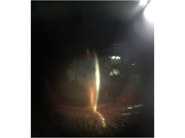
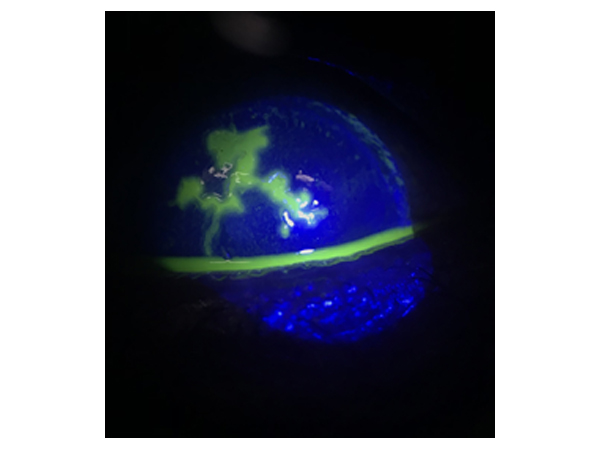
Staining is one of the important tools in ocular examination. This involves application of different dyes as per the requirement and evaluation with viewing system (slit lamp most commonly). Dyes used for this are
Fluorescein: It is the most widely used one. It is an orange dye and best observed under blue light. Whenever there is a defect in the cornea it stains the underlying tissue and appears green on blue light. it helps in
Corneal staining for abrasion, ulcer or keratitis
Dry eye evaluation
Contact lens fitting
Applanation tonometry
Bengal red & Lissamine green: These dyes stain dead cells present on the ocular surface and help in the dry eye evaluation
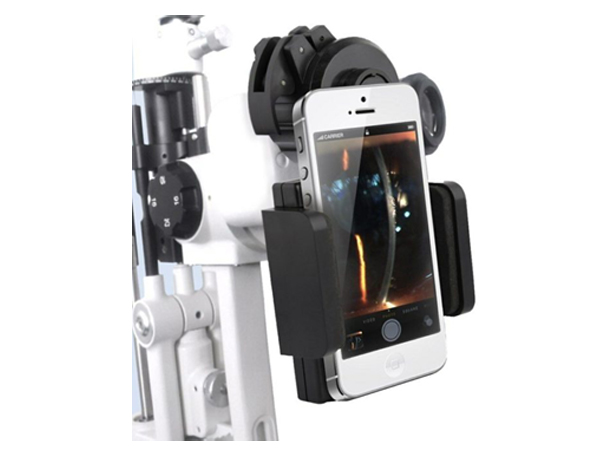
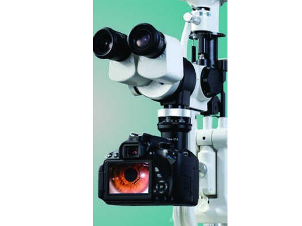
SL Photography
This involves Photographic record keeping of eye lesions with the help of slit lamp and camera