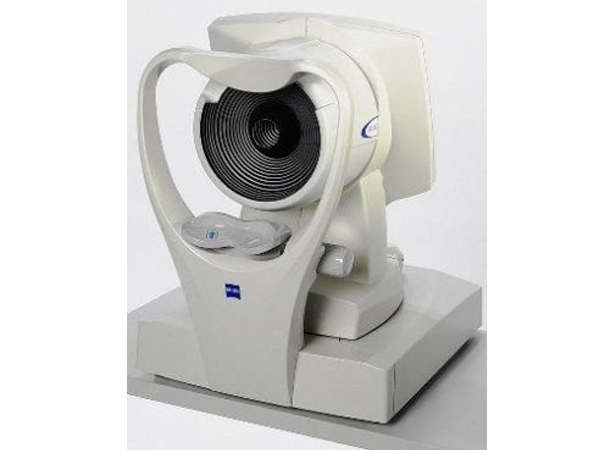
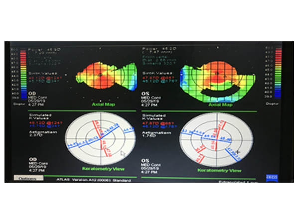
Corneal Topography
Corneal Topography is a non-invasive medical imaging technique for mapping the surface curvature of the cornea, the outer structure of the eye. Since the cornea is normally responsible for some 70% of the eye's refractive power, its topography is of critical importance in determining the quality of vision and corneal health.
Corneal topography is most commonly used for the following purposes
Refractive surgery
Keratoconus
Post surgery astigmatism
Surgical planning in cases with astigmatism
Effect of corneal and ocular surface disorders on tear film
Contact lens fitting
incision placement and intrastromal ring placement in keratoconus
Pachymetry
Corneal pachymetry is the process of measuring the thickness of the cornea. A pachymeter is a device used to measure the thickness of the eye’s cornea. It is used to perform corneal pachymetry prior to refractive surgery, forscreening of Keratoconus, LRI surgery and is useful in screening for patients suspected of developing glaucoma among other uses.


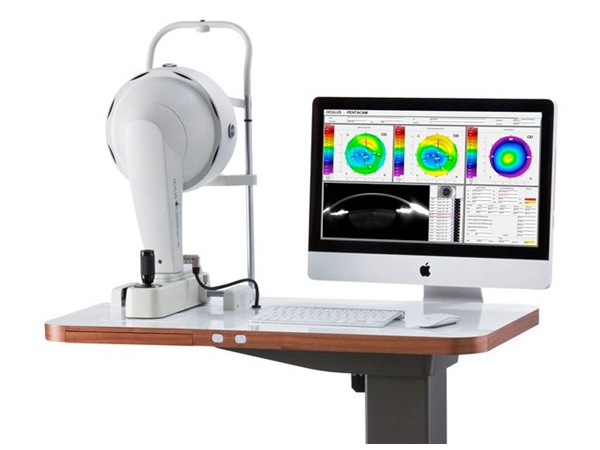
Pentacam
The Pentacam images the anterior segment of the eye by a rotating Scheimpflug camera measurement. This rotating process supplies pictures in three dimensions. The center of the cornea is measured very precisely because of this rotational imaging process. The measurement process lasts less than two seconds, and minute eye movements are captured and corrected simultaneously. By measuring 25,000 true elevation points, precise representation, repeatability and analysis are guaranteed.
The Pentacam provides 3-dimensional chamber analysis of the complete anterior segment of the eye, corneal topography of both the front and the back of the cornea, thickness measurements across the entire diameter of cornea. It also can provide cataract analysis and clearly detect corneal disorders such as Keratoconus, where laser vision correction is sometimes not appropriate.