Schirmer test
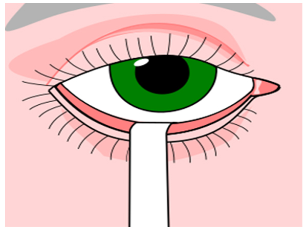
Schirmer test determines whether the eye produces enough tears to keep it moist. This test is used when a person experiences very dry eyes or excessive watering of the eyes. It poses no risk to the subject.
A negative (more than 10 mm of moisture on the filter paper in 5 minutes) test result is normal. Both eyes normally secrete the same amount of tears.
A positive test signifies decreased secretion of tears
Corneal Topography
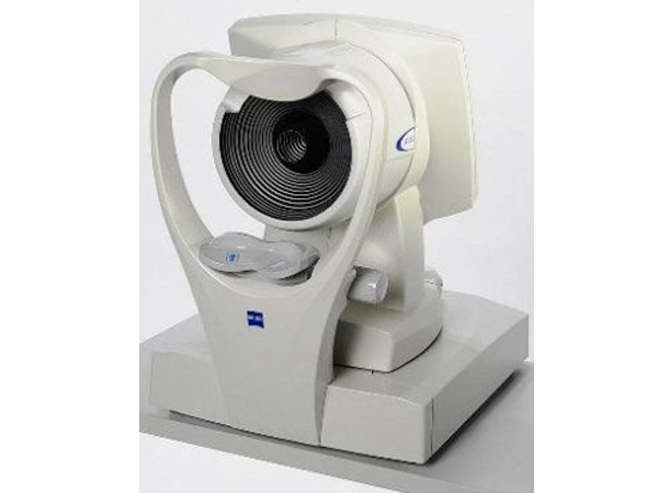
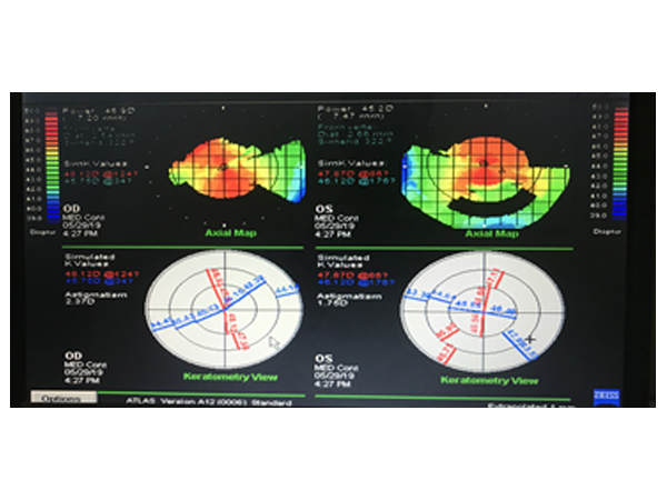
Corneal Topography is a non-invasive medical imaging technique for mapping the surface curvature of the cornea, the outer structure of the eye. Since the cornea is normally responsible for some 70% of the eye's refractive power, its topography is of critical importance in determining the quality of vision and corneal health.
Corneal topography is most commonly used for the following purposes
Refractive surgery
Keratoconus
Post surgery astigmatism
Surgical planning in cases with astigmatism
Effect of corneal and ocular surface disorders on tear film
Contact lens fitting
incision placement and intrastromal ring placement in keratoconus
Tear Film Break Up Time (TBUT)
TBUT is an indication of tear film stability. The proper method of TBUT testing is using a wet fluorescein-impregnated strip. The dye is distributed by blinking, and the patient is then asked to stare straight ahead without blinking. The tear film is observed under the cobalt blue light of a slit lamp, and the time between the last blink and the appearance of the first dry spot or hole in the tear film is measured and equal to the TBUT.
TBUT has been shown to be decreased in keratoconjunctivitis sicca, mucin deficiency, and meibomian gland disease. Normal subjects show variability in TBUT, although 10 seconds is the typical cutoff between normal and abnormal results and has been found to be relatively specific in screening patients for tear film instability.
Corneal Staining
Ocular surface staining is done with dyes and patterns observed. The interpretation guides towards the diagnosis and grading of Ocular Surface Disorders. Dyes commonly used are
Fluorescein sodium
Rose Bengal
Lissamine green
Meibomian Gland Analysis
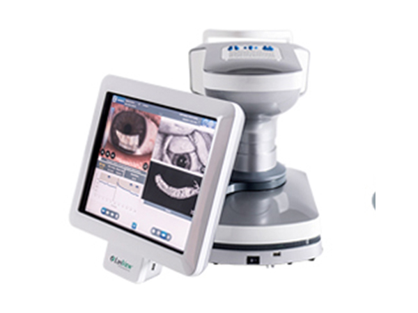
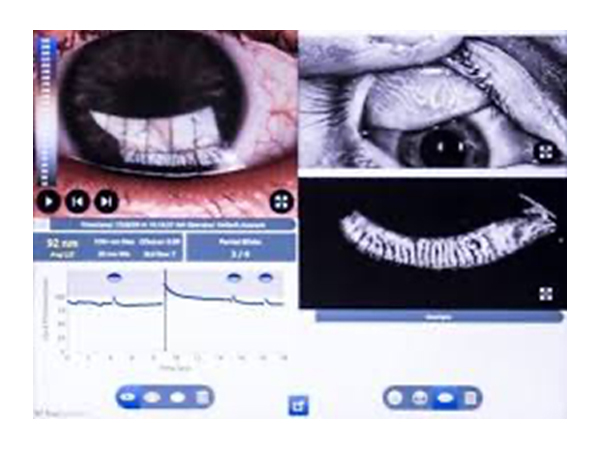
Lipiview is one of the innovative new imaging tools used to detect the level of a patient's dry eyes.
It uses a special digital image of your eye that allows our doctors to see and measure the lipid layer of your tear film. Your doctor is then able to evaluate what, if any, tear therapy would benefit you. It gives structural details of Meibomian glands and help in management of Dry eye
